Minimally invasive inlay restoration from the hybrid ceramic VITA ENAMIC
Inlay restorations using CEREC procedures have been an established process in digital dentistry for decades. However, due to the required minimum wall thickness, a lot of tooth substance frequently had to be dissected in reconstructions of traditional ceramics. Due to reduced minimum wall thicknesses, VITA ENAMIC (VITA Zahnfabrik, Bad Säckingen, Germany) allows minimally invasive restorations and can be precisely ground in thinly tapering edge areas. In the report, Dr. Gerhard Werling (Bellheim, Germany) explains the clinical procedures for an inlay-restoration of hybrid ceramic in region 24-26.
1. Initial situation
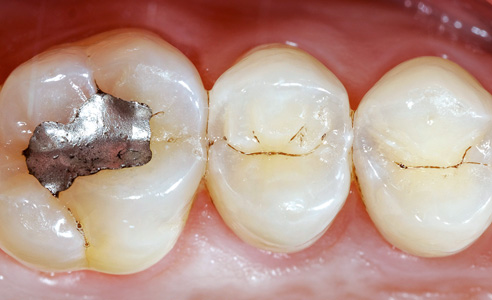
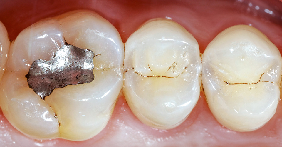
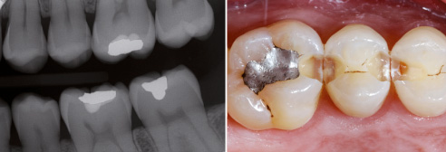
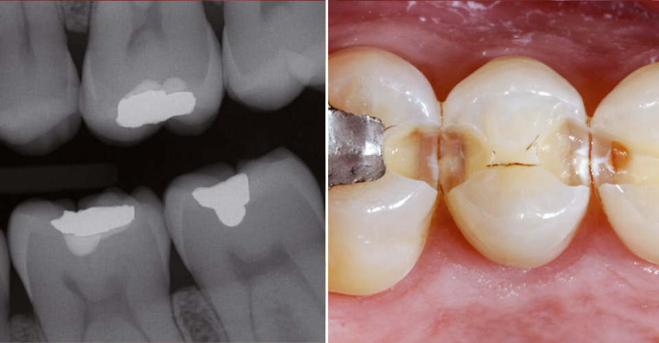
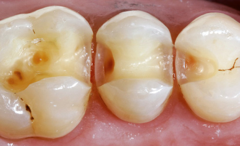
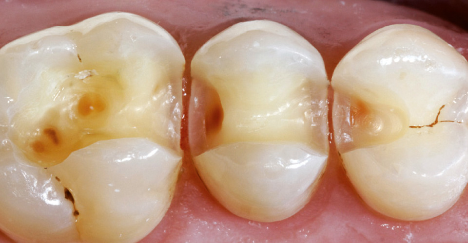
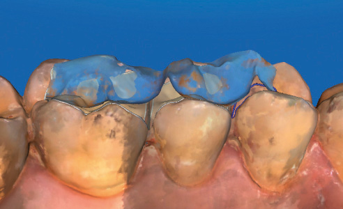
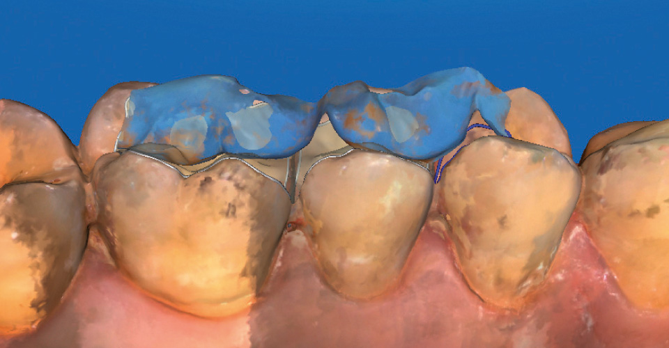
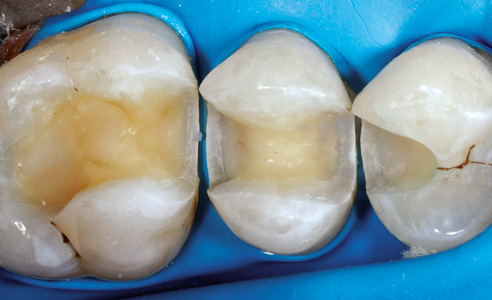
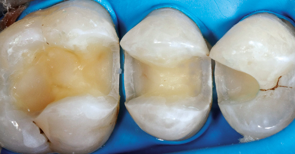
Figures 1 and 2 show the initial situation. On the basis of the patient's history and according to the patient's request (male, 38 years), he was not treated with alternative methods (infiltration technique, fluoridation, regular controls, etc.). Instead, a filling cavity was carefully dissected on the tooth in which the caries had already penetrated the approximal enamel in the X-ray image. Surprisingly, in the clinical image, the caries had penetrated deep into the dentine, so that after extensive excavation, a considerable defect in the substance was present.
2. Material selection
Since the patient wanted a permanent enamel-like and tooth-like restoration, composite could not be used as a restoration material. It was decided to proceed according to the "extension for prevention" rule - but as minimally invasive as possible. The hybrid ceramic VITA ENAMIC is very advantageous in this case. The unique network structure in which ceramic and acrylate polymers interpenetrate, provides for enormous resilience and offers more freedom than traditional restoration materials.
3. CAD/CAM workflow
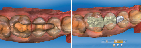
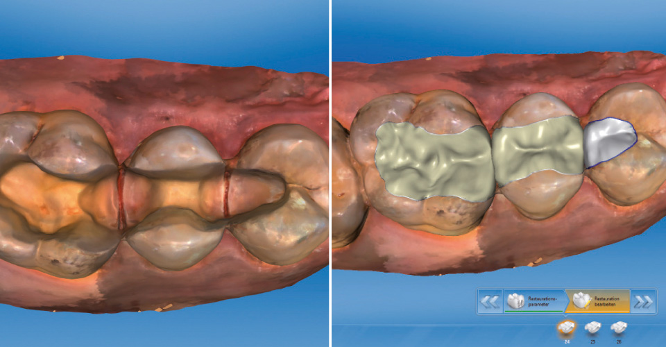
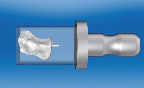
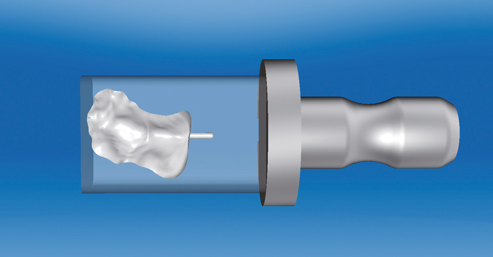
Three VITA ENAMIC inlays were fabricated using the CEREC System (Sirona Dental, Bensheim, Germany). The intraoral scan was done using the CEREC Omnicam. With the biogeneric software, the reconstruction was done analogously to the missing chewing surfaces. In the grinding preview, the inlays were placed in the material blanks. The geometry EM-10 (8 x 10 x 15 mm) was chosen according to the shade determination with VITA Easyshade (VITA Zahnfabrik) in the color 1M2-HT. The hybrid ceramic can be processed very simply and quickly by machine as well as manually. Thanks to the high load-bearing capacity and edge stability, constructions with comparatively small wall thicknesses and thin-running edges are also feasible. Edge chipping, which can occur in traditional ceramics, are rare with this material.
4. Processing and integration
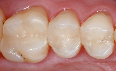
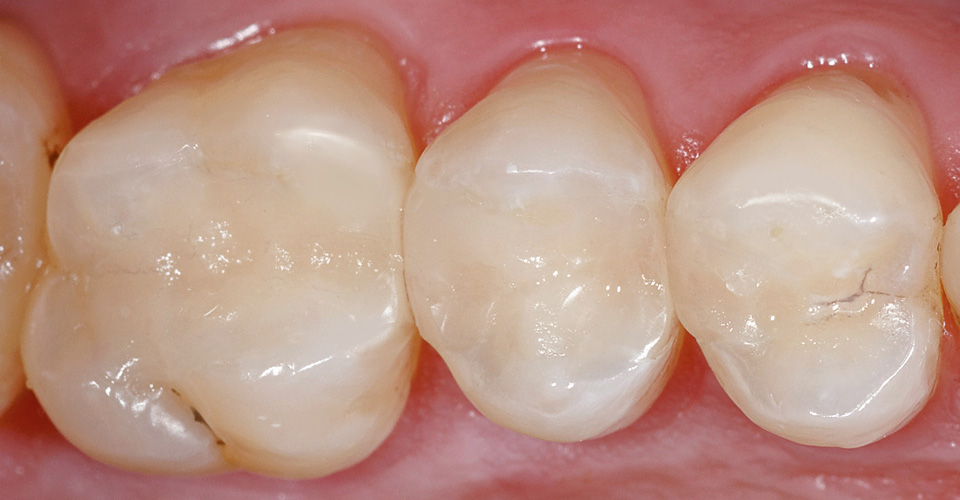
It is advantageous that there is no firing process, and a shade characterization is possible if desired. The available shade selection (0M1 - 4M2) in two translucent steps, plus the good light transmission of the material allow for esthetically pleasing results. The inlays have been polished to a high gloss with the VITA ENAMIC Polishing Set in the clinic. The hybrid ceramic can also be easily polished intraorally. With VITA polishing instruments, the restoration edges can be polished in a unique, fine manner so that virtually no transition between the tooth and the restoration remains visible. Bonding is performed adhesively.
Report 08/16


